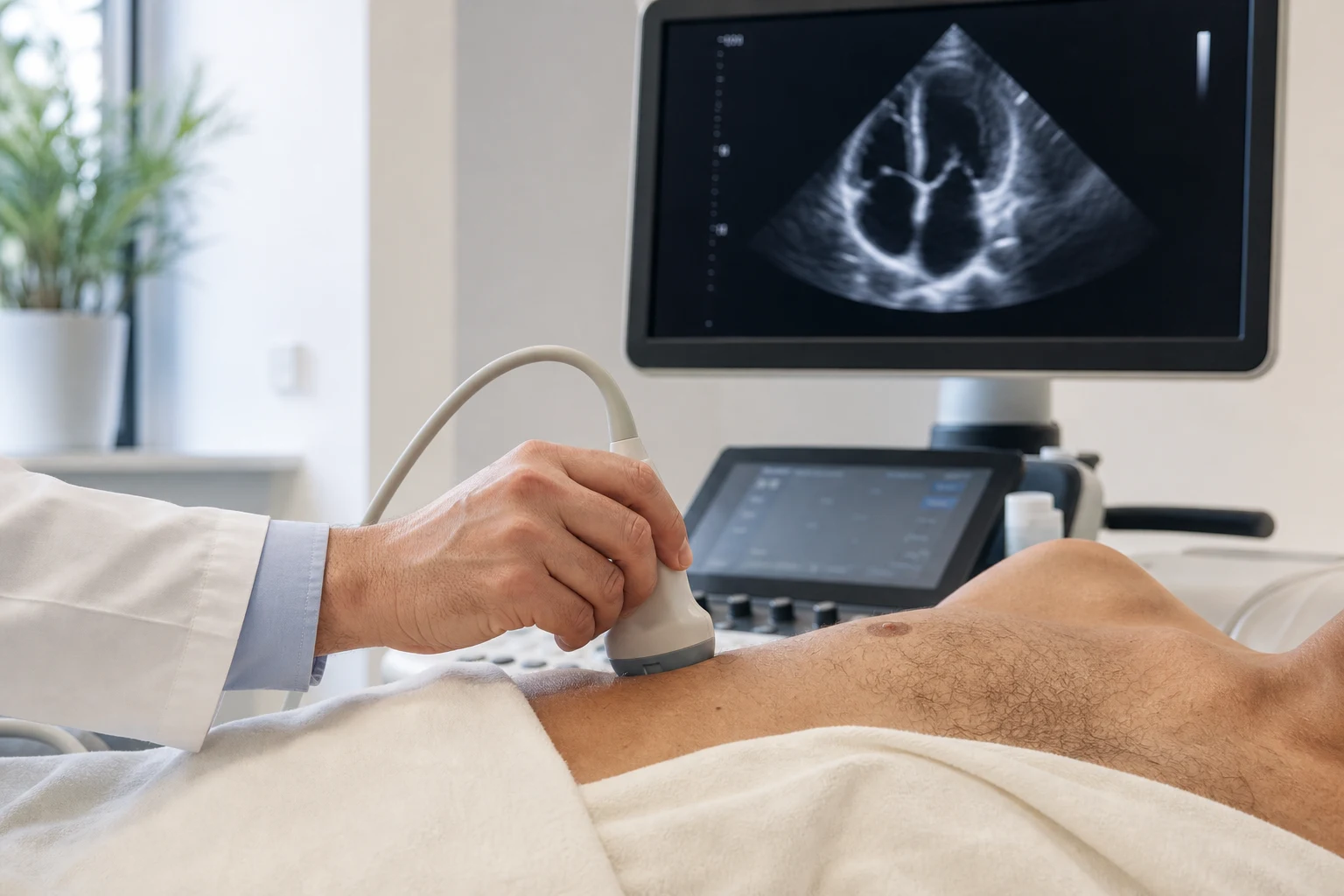
Echocardiogram, or echocardiography, uses ultrasound to visualize the ventricles, valves, contraction, and flow of blood. It is painless and valuable, but a normal result does not rule out, for example, coronary heart disease or intermittent arrhythmia.
At a glance
The main points
- Shows the heart chambers, walls, valves and key measures of heart function.
- It uses no radiation and is painless.
- It does not directly depict the narrowing of the coronary arteries.
- Repeat testing is useful when there is a specific clinical reason or a change in your condition.
How is the exam done?
The examinee usually lies on the left side. With a special head and gel, images are taken from different positions on the chest. Doppler studies the direction and speed of blood flow.
No special preparation is usually required. The quality of the images can be affected by anatomy, breathing or other technical factors, which is why the examination is adapted to each person.
What information can it provide?
The echocardiogram measures the dimensions of the heart chambers, the thickness of the walls and the overall contractile function. It assesses valve movement, possible stenosis or insufficiency, and signs of pressure in the pulmonary circulation.
It can also show fluid around the heart, certain congenital abnormalities, and changes associated with hypertension, heart attack, cardiomyopathy, or heart failure.
What is ejection fraction?
The ejection fraction estimates the percentage of blood ejected by the left ventricle in each systole. It is an important indicator, but it does not describe the entire function of the heart by itself. Heart failure or significant valvular disease may be present even with a preserved ejection fraction.
Small differences between two measurements may also be related to the method or conditions of the test. The trend is interpreted together with the clinical picture.
What doesn't show immediately
A standard echocardiogram does not show the inside of the coronary arteries in detail and cannot, by itself, rule out coronary artery disease. It does not record the heart rhythm for hours as a Holter does, and it cannot replace CT or MRI when a different imaging method is needed.
The choice of test should follow the symptom and the clinical question — not the idea that a “good picture” rules everything.
When is often requested
Indications may be murmur, dyspnea, edema, suspicious ECG, hypertension with possible cardiac effect, known valvular disease, heart failure, or follow-up after a specific cardiovascular event. It is not necessarily needed in every screening.
It repeats every now and then
The interval depends on the finding, severity, symptoms and likelihood of change. A stable or mild picture may require less frequent monitoring, while new dyspnea, decreased strength, or a change in clinical examination may prompt recurrence.
Keep the previous opinion or, ideally, the complete file. Comparing the same metrics over time increases the value of tracking.
Sources & further reading
The article provides general information and is not a substitute for an individualized medical evaluation.